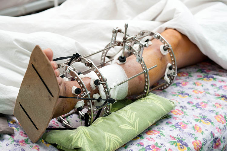
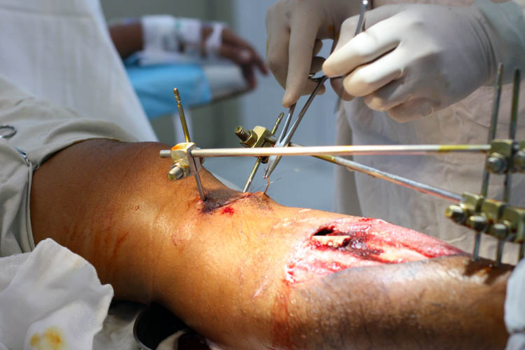
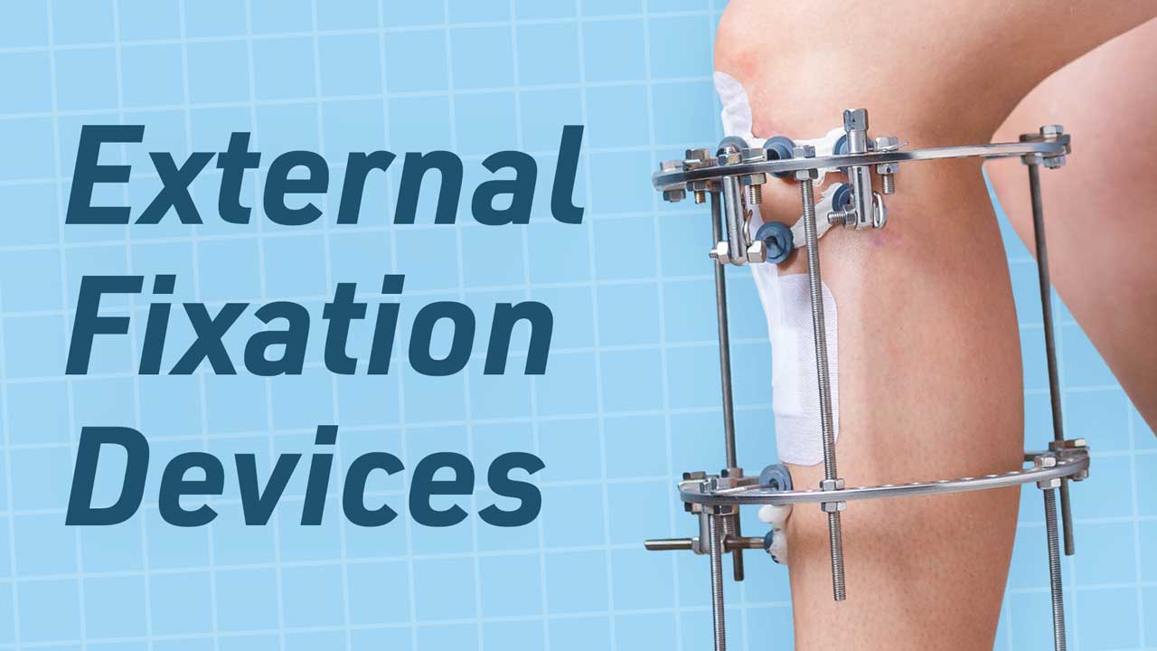
External fixation devices are used to help immobilise a particular part of the body, due to a fracture or certain orthopaedic problem, to allow for bone healing (Singh 2019).
These devices may allow the fixation and manipulation of multiple bone segments, which would otherwise be very difficult. They involve the use of pins, wires and braces, and are used when other options of immobilisation (such as plaster casts) would be ineffective (Singh 2019; Walker 2012).
Fixation devices can be used in limb length discrepancy surgeries, nerve and tendon repairs, and polytrauma patients with fractures, to name a few incidences. They include circular fixators such as halo thoracic braces, Ilizarov fixators, and unilateral fixators (different from circular fixators as they are only positioned on one side of the limb) (Singh 2019).
Risks of External Fixation Devices
There are many risks associated with the use of external fixation devices, including those posed by the device itself, as well as the initial injury that requires fixation.
Pin site complication rates range from 7% to 100%, with the majority of complications being infection. This broad range of numbers is problematic and is due to the lack of a universal classification for pin site infections (Walker 2012).
The risks of external fixation devices include:
- Infection - both pin site and osteomyelitis
- Deep vein thrombosis (DVT) and pulmonary embolisms (PE)
- Aseptic loosening
- Fracture or non-union of existing fracture
- Loss of reduction.
(Roberts et al. 2015; Vaidya et al. 2012)

DVTs and PEs are also potential complications following orthopaedic surgery and are therefore potential complications for those with external fixation devices. However, there is limited evidence that the clots can be caused by the use of the devices themselves. Rather, they are more likely a result of the surgery (Roberts et al. 2015).
Pins within the external fixator can also loosen. This may then create an unstable fixator, which results in an unsuitable environment for bone healing, increased movement in the limb and pin site irritation, which is often a precursor for pin site infection. Pins can loosen for a variety of reasons, one being when the pin is uncoated, which can lead to a fibrous tissue formation where the pin meets the bone (Ferreira & Marais 2012).
Pin Site Infection
One of the most common risks of external fixation devices is infection. A pin site cannot heal whilst the pin and external fixator is in situ. Therefore, it is essential that the pin site is attended to regularly to decrease the risk of potential infection.
One way of looking at pin site wound care is to think of a stab wound: a stab wound can’t heal if the knife is still in the wound. The healing of the site is prevented by the presence of the pin, therefore, wound care revolves around keeping those sites clean and free from infection (Davies et al. 2005).
There are many individual factors that may also increase the risk of the patient developing a pin site infection. These include the patient's age, any pre-existing medical conditions, the reason why they need the external fixation device, and the duration the device is needed for.
A study also found that weather can have an impact on pin site infections, with the rate of infections higher during the warmer seasons (Kao et al. 2015).
The risk of developing a pin site infection increases with the length of time the fixation device is in place. Pin site infections will usually begin as cellulitis, and treatment will depend on the type of infection.
In most cases, a minor superficial infection can be treated with increased pin site care in conjunction with oral antibiotic therapy (Walker 2012). Most infections respond to oral antibiotics, as generally they are caused by a Staphylococcus aureus infection, but sometimes they extend into deeper tissues and bone, causing osteomyelitis, septic arthritis, and in some cases, septicaemia (Walker 2012).
When severe infection occurs, the stability of the fixation can become impaired. This can result in the removal of the pin or wire, but even after its removal, the infection can still linger (Davies et al. 2005). Thankfully, infections are more likely to be superficial than severe. However, even a superficial infection may cause pain and interfere with the patient's recovery and rehabilitation (Davies et al. 2005).
The early recognition of potentially infected pin sites is essential in managing the complication efficiently. This involves documentation and monitoring of all pin sites through regular pin site care. Staff and patients should take particular note of the presence and extent of erythema, tenderness, swelling and discharge (Walker 2012).

Pin Site Care
There is little evidence to support one type of pin care protocol over another, and this can be attributed to the fact that there is no validated grading system or definition for pin site infections (Lee et al. 2012).
Some protocols involve the use of antiseptic solutions, while others use pressure dressings to restrict the movement between the skin and the pin (Davies et al. 2005). The use of pressure dressings can be especially beneficial to pins sites that are near joints, which tend to be more prone to infection due to increased amounts of movement (Davies et al. 2005).
Due to a lack of clear evidence, there are many inconsistencies with pin site management and prevention of pin site infections. However, the goal of management should be to prevent the colonisation of the pins and wires, and therefore, prevent infection (Walker 2012) with regular pin site care.
A popular method of pin site care involves using normal saline or an antimicrobial agent and gauze to clean the pin site areas. This can be done twice a day, daily or even weekly depending on protocols (Lee et al. 2012).
Pin site care protocols are dependent on a variety of factors and are often different depending on the preferences of the surgeon and staff, habit, consensus and basic principles of wound care (Davies et al. 2005). Keep in mind that complete healing of the site is not the goal of pin site care, so some wound care techniques may be inappropriate (Davies et al. 2005).
Staff must also ensure that patients are educated on the signs and symptoms of infection so that complications can be monitored. Patients must also be educated about the restrictions enforced on them due to both their injury or surgery and the use of the external fixation device, for example, being non-weight bearing through the affected limb. It is also important to elevate the limb postoperatively and whenever the patient is not mobilising. This will help reduce oedema around the pins, and therefore, improve the environment around the pin sites (Ferreira & Marais 2012).
Conclusion
External fixation devices come with many risks and benefits to the patient. Due to a lack of clear consensuses, pin site infections and protocols lack much reliable evidence. Therefore, many protocols and practices will vary depending on a variety of factors. It is important that any care given to the patient with an external fixator device is individualized to that person and their injuries.
Topics
References
- Davies R, Nayagam, S & Holt, N 2005, 'The Care of Pin Sites with External Fixation', The Journal of Bone and Joint Surgery, vol. 87, viewed 12 October 2023, https://www.researchgate.net/publication/7883392_The_care_of_pin_sites_with_external_fixation
- Ferreira, N & Marais, LC 2012, 'Prevention and Management of External Fixator Pin Track Sepsis', Strategies in Trauma and Limb Reconstruction, vol. 7 no. 2, viewed 12 October 2023, https://link.springer.com/article/10.1007/s11751-012-0139-2
- Kao, HK, Chen, MC, Lee, WC, Yang, WE & Chang, CH 2015, 'Seasonal Temperature and Pin Site Care Regimen Affect the Incidence of Pin Site Infection in Pediatric Supracondylar Humeral Fractures', BioMed Research International, viewed 12 October 2023, https://www.hindawi.com/journals/bmri/2015/838913/
- Lee, CK, Chua, YP & Saw, A 2012, 'Antimicrobial Gauze as a Dressing Reduces Pin Site Infection', Clinical Orthopaedics and Related Research, viewed 12 October 2023, https://pubmed.ncbi.nlm.nih.gov/21842299/
- Roberts, JS, Panagiotidou, A, Sewell, M, Calder, P & Goodier, D 2015, 'The Incidence of Deep Vein Thrombosis and Pulmonary Embolism with the Elective Use of External Fixators,' Strategies in Trauma and Limb Reconstruction, vol. 10, no. 2, viewed 12 October 2023, https://link.springer.com/article/10.1007/s11751-015-0219-1
- Singh, AP 2019, 'External Fixation Devices – Concept and Use', Bone and Spine, 2 August, viewed 12 October 2023, https://boneandspine.com/external-fixation-devices/
- Vaidya, R et al. 2012, 'Complications of Anterior Subcutaneous Internal Fixation for Unstable Pelvis Fractures: A Multicenter Study', Clinical Orthopaedics and Related Research, vol. 470, no. 8, viewed 12 October 2023, https://pubmed.ncbi.nlm.nih.gov/22219004/
- Walker, J 2012, 'The Problem with Pin Site Infection', Journal of Nursing and Care, viewed 12 October 2023, https://www.hilarispublisher.com/open-access/the-problem-with-pin-site-infection-2167-1168.1000e111.pdf
 New
New 
